
Why we should know about stimulation threshold of D wave.
Its important to know D wave is play important role in specially in Intramedullary spinal cord tumors , to assess the continuity of spinal cord functions while decompressing the tumor from inside the cord. this tumors that grow within the spinal cord itself, presenting unique challenges during surgery due to their location and potential impact on neural function.
Stimulation Intensity of D wave.
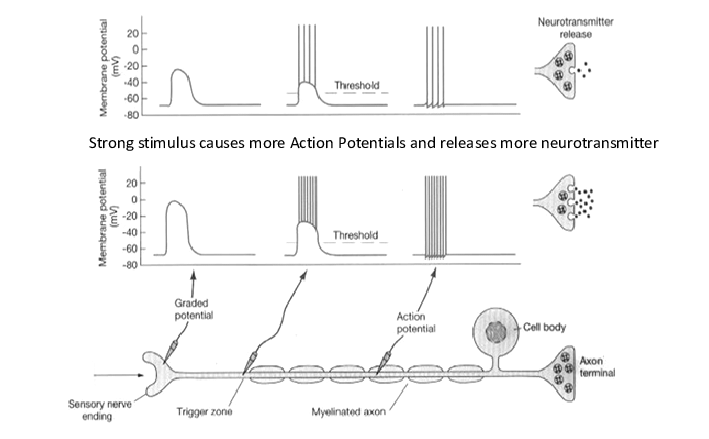
- Transcranial electrical stimulation (TES) is critical. It should be sufficient to depolarize the corticospinal tract (CST) neurons responsible for generating the D-wave response.
- However, caution must be exercised to avoid excessive stimulation intensity that could lead to unwanted side effects like tongue laceration , disturbance of surgical field because its a average potentials or neural injury.
Location of stimulating electrodes for D wave.
- For stimulation site should be over the motor cortex corresponding to the muscles innervated by the spinal cord segment affected by the tumor.
Stimulation Technique.
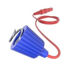
- TES is commonly used for D-wave monitoring, delivering electrical pulses to the motor cortex invasively though corkscrew electrode.
Type of Pulse and Frequency for D wave.
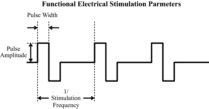
- Non deposition of charge biphasic pulses are often preferred for their effectiveness and safety in eliciting D-wave responses.
- The frequency of stimulation pulses can vary depending on the specific protocol and equipment used, typically ranging from single pulses to bursts of stimuli.
Monitoring and Adjustment.

- During procedure , continuous monitoring of D-wave responses is performed to assess the real-time integrity of the corticospinal tract
- Stimulating parameters may be adjusted based on the observed responses, like amplitude their latency .
Stimulating parameter.
As per standard practice through text book , A single pulse along 50-75 microsecond pulse duration at the rate of 1Hz , intensity of current usually keep 80-90 Volt or this parameter may be adjusted through clinician .
Recording Parameters.
Placement of Electrode.
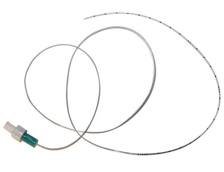
- Epidural platinum electrode usually to be placed at caudal to surgical side and if possible other electrode to be placed rostrally as a control.
Filter Settings.
- Filters are used to isolate and amplify the signals of interest while reducing noise. Common filter settings include a bandpass filter with cutoff frequencies between 30 Hz and 15 kHz. or it can be changed according to signal quality.
Averaging of Signal.
- Multiple stimuli are often delivered and averaged to improve the signal-to-noise ratio and enhance the visibility of D-wave responses.
Display Parameters.
- The recorded signals are displayed on a r computer screen, where technologist and clinicians can analyze the waveforms for amplitude, latency, morphology, and other characteristics. usually we keep 2ms /division total sweep speed 20 millisecond because its contains shorter latent period .
Conclusion.
- This is very much important to understand that the stimulating parameters may vary slightly depending on the specific surgical context, patient factors, equipment availability, and institutional protocols. surgical electrophysiologist and surgical teams work closely together to tailor these parameters and ensure the effectiveness and safety of D-wave monitoring during intramedullary spine tumor surgeries.
Related to this article.
https://neurointraoperative.com/wp-admin/post.php?post=1465&action=edit
https://www.ncbi.nlm.nih.gov/pmc/articles/PMC3631052
Question.
- How much current threshold need to get the D wave potentials?.
- Why we record D wave as a average potentials ?.
- D wave is a motor response?.
- Where is the placement of D wave electrode in spinal cord?.
- Where is the placement of corkscrew electrode for stimulation?.
1 thought on “D Wave Stimulating and Recording .”