
- Electrical Stimulation induced seizures as a modality/tool of mapping the ictal onset zone. Stimulation can be performed at 1 Hz for 20–40 second or at 50 Hz at maximum 5 second duration . Note High frequency for short duration and less frequency for a little long time.
Electrical stimulation seizure site.
- The target of the brain where the electrical stimulation being applied is crucial in inducing a seizure. The stimulation site will be selected based on preoperative imaging and as per based on routine or video EEG findings along with seizure semiology.

Amplitude of electrical Stimulation .
- The strength of the electrical stimulation is measured in milliamperes (mA) and it is gradually increased to a level sufficient to induce a epileptic electrographic discharges . The intensity stimulation will vary based on individual subject/patient characteristics and the response to stimulation.
Pulses of stimulation .
- The frequency at which the electrical pulses are delivered and it is influence the likelihood of inducing a epileptic electrographic discharges
- Parameters such as single stimulations pulse , trains of stimuli, or high-frequency stimulation may be used depending on the clinical outcome.
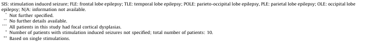
Summary of studies on stimulation provoked/induced seizures.

Monitoring and Safety .
- During electrical stimulation provoked epileptic electrographic discharges, continuous monitoring of the patient’s vital signs, invasive EEG activity, and seizure semiology is crucial to ensure patient safety. Emergency medications and equipment should be readily available to manage any adverse events.
summary.
- Electrical Stimulation-provoked epileptic electrographic discharges are a valuable tool in before elective epilepsy surgery to help identify the epileptic focus and map functional brain areas. And also helps to manage the time from the spontaneous long recording invasive electroencephalography.
- Stereo-EEG plays a prominent role in the presurgical evaluation of focal epilepsies also in the first years of life and that it may offer a surgical option in particularly complex cases that would have scarcely benefitted from further medical treatment.
- Stereoelectroencephalography monitoring is a helpful and well-tolerated technique for the definition of the epileptogenic zone in complex cases of childhood drug-resistant epilepsy. It provides essential information for safe resections that should result in excellent outcomes in a considerable number of patients.
- SEEG is a safe and highly accurate method that provides essential guidance for epilepsy surgery. Implementing SEEG in conjunction with multimodal planning systems and robotic devices can further increase safety margin, surgical efficiency, and accuracy.
- Robot-guided sEEG electrode implantation using CT-frame referencing and CT-laser-based referencing is most accurate and can serve for high precision placement of electrodes. In contrast, 3.0-T MRI-laser-based referencing is less accurate, but saves radiation. Most trajectories can be reached if alternative routes over less vascularized brain areas are used. This article is part of the Special Issue “Individualized Epilepsy Management: Medicines, Surgery and Beyond.
- SEEG is a safe and accurate procedure for the invasive assessment of the epileptogenic zone. Traditional Talairach methodology, implemented by multimodal planning and robot-assisted surgery, allows direct electrical recording from superficial and deep-seated brain structures, providing essential information in the most complex cases of drug-resistant epilepsy.
- This standardized naming convention, Standardized Electrode Nomenclature for SEEG Application, provides a simple, concise, reproducible, and informative method for specifying the target(s) and relative position of each SEEG electrode in each patient, allowing for successful sharing of information in both the clinical and research settings. General adoption of this nomenclature could pave the way for improved communication and collaboration between institutions.
- SEEG is a safe and effective technique for invasive SOZ localization in medically refractory LRE in the pediatric population. SEEG permits bilateral and multilobar investigations while avoiding large craniotomies. It is conducive to deep, 3D, and perilesional investigations, particularly in cases of prior resections. Patients who are not found to have focally localizable seizures are spared craniotomies.
REF-

Related this article.
https://neurointraoperative.com/wp-admin/post.php?post=1919&action=edit
1 thought on “Parameters Direct Electrical Cortical Stimulation Induced Seizures.”