
Historical perspective.
- Direct Cortical Stimulation was used to define the epileptogenic zone in patients undergoing invasive pre-surgical investigations with depth electrodes for pharmacoresistant epilepsy {Bancaud et al.,1974}.
Introduction of seizures provoked by direct Cortical Stimulation.
- In Cortical Stimulation provoked seizures the Invasive Electroencephalography[EEG] discharges are triggered as an immediate result of the electrical stimulus see in figure I.
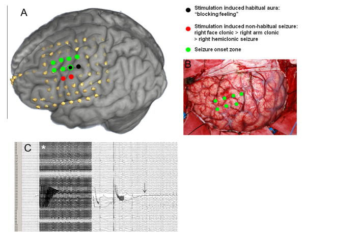
- Illustration of spontaneous and stimulation induced seizure onset zones. [A] Co-registration of the electrode positions with the patient’s preoperative MRI. Electrodes involved in the stimulation induced ‘‘habitual’’ affected onset are highlighted in black, those involved in non-habitual stimulation induced seizure onsets are highlighted in red.
- Electrodes involved in the seizure onset zone of spontaneous seizures are highlighted in green. Note that the three areas are in close proximity, but not completely concordant.
- [B] The photograph shows the exposed cortex in the operating room; green dots highlight the area of the affected onset zone, where electrodes were positioned previously.
- [C] Intracranial EEG recording of a non-habitual stimulation induced seizure. The white asterisk marks the onset of the stimulation artefact. Note that after discontinuation of the stimulation, the after discharge develops at the stimulation site (black arrow) and then evolves into a electrographic pattern with spread to adjacent electrodes. The semiology of this epileptic electrographic was non-habitual starting with a right face clonic seizure evolving into a right arm and subsequently right hemiclonic electrographic .
- If symptom is generated during the stimulation before significant propagation of the electrical discharge has occurred which means that there is close co-localization of a cortical region that can generate this symptom and can be considered as the symptomatogenic area of the Cortical Stimulation.
Aspect of Cortical Stimulation provoked seizures in presurgical epilepsy examination .
- By use of Cortical Stimulation induced seizure to define eloquent cortex. Eloquent cortex refers to areas of the brain that are involved in critical functions such as movement, sensation, language, and cognition. Identifying these regions is crucial in neurosurgery, as they must be preserved to avoid postoperative deficits. Note if involvement onset zone localized within these functional areas of the brain, there is role of team primary to be confirmed , through STEREO EEG recording electrode with ozomen/Wilder Penfield stimulation technique, {invasive recording electrode can be used as a stimulator } during stimulation and just after stimulation electrographic irritating zone if confirms in these functional areas, the surgical technique is call disconnection means isolate the seizure originating gyrus , for example if source is motor area so that precentral gyrus will disconnect from post central gyrus or adjustant areas if its require, because if surgeon resects epileptogenic tissue from the motor area patient can reveal with motor deficits .
Stimulation induced seizures which together with the interpretation of the spontaneous seizure define the epileptogenic zone Figure II.
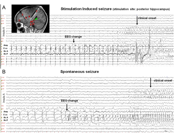
- Figure II Stimulation induced versus spontaneous epileptic electrographic . Intracranial stereo-EEG (multiple depth EEG electrodes) traces showing a stimulation induced electrographic (induced by 1 Hz electrical stimulation.
- [A] and a spontaneous seizure [B] in the same patient. Insert in panel A shows an MRI with the position of the stereo-EEG electrodes. A green flash marks the stimulation site. Note that the stimulation induced epileptic electrographic [A] and the spontaneous seizure [B] have similar EEG characteristics and both show spread into the insular region coinciding with the clinical epileptic electrographic onset.
Stimulating settings to provoked seizures.
- The stimulation [parameters] that influence the likelihood of inducing a epileptic electrographic through stimulation will vary depending on the individual’s medical history, the specific condition being targeted for elective epilepsy surgery whoever not responding with antiepileptic drugs{AEDs}
Type of Stimulation.
- The electrical stimulation being used and its significantly enhance epileptiform activity like spike wave discharged type of morphology while does occur in spontaneous stage but indefinite or time taking , and also target the exact location of origin of epileptic electrographic so that while procedure being planned to remove the epileptic zone that means during surgery adjacent normal brain tissue should not get affect. [Note-at direct CS induced epileptic electrographic as a tool of mapping the ictal onset zone].
Strength of stimulus.
- The intensity of the stimulation refers to the strength of the stimulus applied. Higher intensity stimulation may be more likely to induce a electrographic compared to lower intensity stimulation. And amplitude of stimulation its vary / will vary on individual subject who targeting for surgery.
Number of pulses in one second [Frequency]
- The number of pulses of the stimulation refers to how often the stimulus is applied within a given time period. Certain frequencies of stimulation pulses may have a higher propensity to induce seizures in susceptible individuals.
Time interval [Duration]
- The duration of the stimulation refers to how long the stimulus is applied each time. Prolonged stimulation may increase the risk of inducing a epileptic electrographic compared to shorter durations. Time interval will vary according to prognostication.
Area.
- The specific brain region being targeted for stimulation, usually its influence the cortical stimulation induce epileptic electrographic discharges.
- Analysis of waveform morphology in invasive electroencephalography.
- Close monitoring of recording side during stimulation is crucial for detecting any signs of epileptic electrographic activity promptly.
Titration
- Gradually increasing the stimulation parameters over time (titration) can help identify the threshold at which seizures are more likely to occur while minimizing the risk of, for a long time electrographic and clinical seizure.
Brief about stimulation and recording.
SEEG is a minimally invasive procedure. It uses electrodes placed directly in the brain to identify where epileptic seizures start.

By use of Stereo-electroencephalography electrode intraoperative stimulation to induce seizures and as well as recording with adjacent electrode.
Most of the centers uses a high frequency stimulation in the range of 50–60 Hz, intensities along with range of 0.25–15 milliampere.
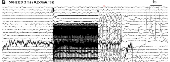
B 50 Hz electrical stimulation [ES]) over 4 second. The onset of the stimulus artefact is marked with an open arrow and the offset with a solid arrow. A seizure pattern is seen outlasting the stimulation artefact.
conclusion.
By cortical stimulation provoked Seizure its appears to identify the epileptic generator as reliably as spontaneous seizures, purpose of this latest technique lead to a more time-efficient intracranial pre-surgical investigation of epilepsy as the need to record spontaneous seizures is reduced.
Related this article.
https://neurointraoperative.com/wp-admin/post.php?post=1931&action=edit
https://pubmed.ncbi.nlm.nih.gov/31180505
Note – In a few place, seizure word used as a epileptic electrographic due to my channel optimization .
Question.
Q-Why we use stimulation induced seizure for surgical candidate?.
A-To localize the actual source of epileptic zone
Q-Is really this technique useful from the spontaneous EEG?
A- In some cases when routine EEG/VEEG findings are not clear{semiology}
Q-How this technology can reduced the timing of long recording EEG?.
A- purpose of this latest technique lead to a more time-efficient intracranial pre-surgical investigation.
Q-How to interpret the EEG trace, while stimulation being done?.
A– while stimulation being done we see which electrode firing much{spike} and from which location of brain.
This website online is really a stroll-by way of for all the data you wished about this and didn’t know who to ask. Glimpse right here, and also you’ll undoubtedly discover it.