
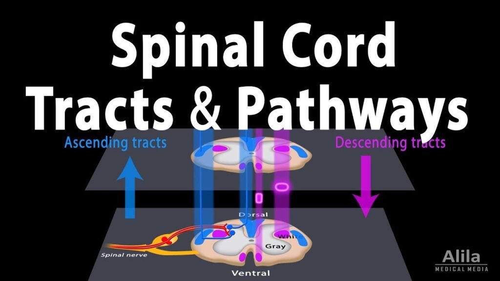
Introduction of motor system {Descending tract}.
Generally an injury to the spinal cord will be most likely be picked up by somatosensory evoked potentials (SSEPs), yet a focal injury to the anterior spinal artery (ASA) may be missed There is a lot of evidence in the literature describing selective injury to the anterolateral columns sparing dorsal columns with preserved SSEPs .
Iatrogenic injuries are an undesired consequence of surgery, yet iatrogenic injuries to the Descending tract system are much more devastating to a patient’s quality of life than most injuries to the sensory
system.
The inclusions of motor evoked potentials (MEPs) to the intraoperative monitoring toolbox can help to confirm/prevent selective lesions to the anterolateral columns of the spinal cord. Yet MEPs are not without their limitations. Even with these limitations, proper application and interpretation of MEP data can be a significant adjunct in reducing iatrogenic injury during surgery.
The anatomy involved in generating MEPs includes various structures within the central nervous system (CNS) and peripheral nervous system (PNS). Here's an overview of the key anatomical components related to MEPs:
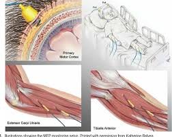
Pre-central Gyrus {Cortex}.
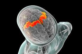
- The Pre-central Gyrus cortex is located in the precentral gyrus of the frontal lobe, specifically in Brodmann areas 4 and 6. It is responsible for planning, initiating, and controlling voluntary movements.
- Stimulation of the Pre-central Gyrus cortex is typically achieved using techniques like transcranial electrical stimulation (TES) or direct electrical stimulation .
Upper Motor Neurons (UMNs).
- UMNs are located within the Pre-central Gyrus cortex and the descending tract pathways, including the corticospinal tract.
- Corticospinal neurons originate in the Pre-central Gyrus cortex, descend through the internal capsule, and travel down the brainstem (pyramid of the medulla oblongata) to synapse with lower motor neurons in the spinal cord and brainstem nuclei.
Corticospinal Tract.
- The corticospinal tract is a major descending tract pathway responsible for voluntary movement control.
- It consists of two main divisions: the lateral corticospinal tract (controls limb muscles) and the anterior corticospinal tract (controls axial and girdle muscles).
- Fibers from the corticospinal tract synapse with lower motor neurons in the anterior horn of the spinal cord gray matter.
Lower Motor Neurons (LMNs):
- LMNs are located in the anterior horn of the spinal cord gray matter (ventral horn) and brainstem motor nuclei.
- They receive input from UMNs and relay motor commands to the skeletal muscles via peripheral nerves.
Peripheral Nerves.
- Peripheral nerves carry Descending tract signals from the spinal cord and brainstem to the target muscles.
- The nerves innervating specific muscles are responsible for transmitting the voluntary commands that lead to muscle contraction.
Muscles.
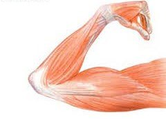
- Skeletal muscles are the effector organs of the Descending tract system, responsible for generating movement in response to motor commands.
- MEPs are recorded by placing surface electromyography (EMG) electrodes on specific muscles, typically in the limbs, to detect muscle responses following motor cortex stimulation.
EMG Recording.
- EMG electrodes are used to record the electrical activity generated by muscle contractions in response to Descending tract commands.
- The EMG signals are amplified, filtered, and processed to isolate the MEPs and assess their characteristics, such as amplitude, latency, and waveform morphology.
Information about the integrity and function of the central and peripheral Descending tract pathways. Here's a detailed look at the physiology of Descending tract Evoked Potentials.
Descending tract Pre-central Gyrus Cortex Stimulation:
- MEPs are typically elicited by stimulating the Pre-central Gyrus cortex, specifically the primary cortex located in the precentral gyrus of the frontal lobe.
- Transcranial electrical stimulation (TES) is a common method used to stimulate the Descending tract cortex invasively. It involves applying a brief, strong magnetic field to the scalp, which induces electrical currents in the underlying cortical neurons.
Upper Motor Neurons (UMNs).
- When the Descending tract cortex is stimulated, it activates the upper motor neurons (UMNs) located in the cortex and the descending motor pathways, particularly the corticospinal tract.
- UMNs transmit motor signals from the cortex to the lower motor neurons (LMNs) located in the spinal cord or brainstem.
Corticospinal Tract Activation.
- The corticospinal tract is a major descending pathway that controls voluntary movements. It consists of fibers originating from the motor cortex, which descend through the internal capsule, brainstem (pyramid of the medulla oblongata), and terminate in the spinal cord.
- Stimulation of the Descending tract cortex or the corticospinal tract directly activates these descending fibers.
Lower Motor Neurons (LMNs).
- In the spinal cord, the corticospinal tract synapses with lower motor neurons (LMNs) in the anterior horn of the gray matter.
- LMNs are the final common pathway for motor signals. They send voluntary commands to the skeletal muscles through peripheral nerves, leading to muscle contraction.
Muscle Response.
- When the LMNs receive Descending tract signals, they depolarize and generate action potentials that propagate along the peripheral nerves to the target muscles.
- The muscle fibers then contract in response to the motor commands, generating electrical activity known as electromyographic (EMG) signals.
MEP Recording.
- EMG electrodes are placed on specific muscles of interest, typically in the limbs, to record the muscle responses.
- The EMG signals generated by muscle contractions are amplified, filtered, and processed to isolate the MEPs from background noise.
MEP Waveform Analysis:
- The MEP waveform includes components such as amplitude, latency, duration, and morphology.
- Neurophysiologists analyze these parameters to assess the integrity, excitability, and conduction velocity of the motor pathways.
- Changes in MEP characteristics can indicate abnormalities such as central nervous system lesions, spinal cord injury
Summary.
MEPs reflect the functional integrity and connectivity of the descending tract pathways, including the motor cortex, corticospinal tract, lower motor neurons, peripheral nerves, and muscles.
Motor Evoked Potentials (MEPs) are neurophysiological responses elicited by stimulating the Pre-central Gyrus cortex or Descending tract pathways. The anatomy involved in generating MEPs includes various structures within the central nervous system (CNS) and peripheral nervous system (PNS).
Note- This is short article about of Descending tract Evoked potential for theory purpose .
{All above theory will explain in details}.
Related to this article.
https://neurointraoperative.com/wp-admin/post.php?post=136&action=edit
https://www.e-neurospine.org/journal/view.php?number=265
Question
- Why we should know about descending tract?.
- Which fibers we stimulate and record in MEP?.
- Which Gyrus we stimulate in descending tract Evoked Potentials, Pre-central or post central Gyrus?.
- What kind of waveform morphology we record in MEP, polyphasic/monophasic?
- Which types of potentials we get in MEP, myogenic/neurogenic?.
1 thought on “Theory of Transcranial Motor Evoked potential MEP.”