What are the indications for functional speech mapping.
- Intracranial glioma/pathology specially in the language-eloquent areas of the brain, can affect one’s speaking ability. Despite importance in surgery, the excision of these lesions can be challenging. Intraoperative neurophysiological monitoring (IONM) during awake craniotomies can help identify language eloquent areas and minimize postoperative impairments. Preoperative language testing is mandatory to establish a baseline before intraoperative language testing.
Technical setups.
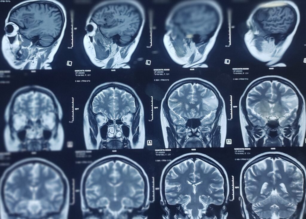
Magnetic Resonance Imaging (MRI) and Diffusion Tensor Imaging (DTI).
Preoperatively several preparations must be taken to ensure that direct cortical stimulation (DCS).
Lesion near to Broca’s area.

Perioperative task during surgery.
Speech/Language Tasks
- Direct Electrical cortical/subcortical Stimulation carried out clinically are performed to locate language areas in the dominant hemisphere. A set of common language mapping tasks (counting, picture naming, reading) can be to activate those area as well speech arrest will observed with these tasks varying according to brain tumors.
- Electrical Stimulation: Surgeons will use electrical stimulation to temporarily disrupt brain function in specific areas to see if it affects the patient’s speech. This helps identify which areas are critical for language and should be avoided during the procedure.
Number Counting and visual task.

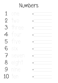
- Counting number like 1-10 is an automatic speech task that is widely being used in language assessment especially in patients who cannot complete complex cognitive tasks.
- While language mapping being assessed during procedure , number counting is carried out first to identify cortical and subcortical structures related to speech output.
Picture Naming when lesion near to comprehensive area.
Lesion on the Wernicke’s area.

Picture naming is one of the most widely being used tasks in language mapping. { like Banana, Apple, phone, car etc}.


Stimulating Parameters.
- Single square pulse at the rate of {@} 60-50 Hz {Penfield/Ozome}.
- Pulse duration- 1 millisecond/1000 microsecond.
- Current intensity will vary according to subject like any language related deficit etc.
- Usually starts 3-5 milliampere.{for short epochs 3 to 8 seconds in duration}.
Anesthesia.
- Local/Scalp block.
Conclusion.
- Accurate techniques with reliable identification of the mapping thresholds are essential for avoiding permanent language related neurologic deficits.
Related to this article.
https://neurointraoperative.com/wp-admin/post.php?post=2204&action=edit
https://www.frontiersin.org/articles/10.3389/fnhum.2021.612891
Question.
- Q-Can we do language mapping in general anesthesia?.
- A-Language mapping is a subjective test which means during perioperative time the patient has to follow the command like verbal fluency, object naming etc.
- Q-Broca’s Mapping is possible under sedation like tongue twist or lip movement?.
- A-literature says if tumor in pure Broca’s area it is possible to see lip movement , tongue twist but other possibility of limbs and face and neck movement because motor speech fibers are close to motor strip posterior wall.
- Q-Is possible functional speech mapping in every patient like lesion on dominating side?.
- A- in every subject functional speech mapping would not be possible because other possibilities are , like age factor , psychometric assessment if not clear, epilepsy patients , Difficulty in to understand the task or commands etc.
- Q-Subcortical speech fibers also should map while resecting tumors from these are?.
- A-Yes absolutely should mapped subcortical speech fibers as well because if these functional neural elements are gone during resection of tumors which means permanent speech related neurological deficits can occur.
- Preoperative Planning: Before surgery, imaging studies like MRI or fMRI are used to identify key language areas. Surgeons also review the patient’s language abilities to establish a baseline.
- Mapping and Preservation– By combining the results from speech tasks and electrical stimulation, surgeons can create a detailed map of the brain’s language regions. This guide helps them navigate around these areas to minimize the risk of postoperative speech deficits.
- Intraoperative Monitoring– During surgery, the patient is usually awake and can participate in tasks like naming objects, repeating words, or reading sentences. This real-time testing helps map out the brain regions involved in these tasks.

1 thought on “What are the Stimulating parameters for speech mapping.”