

During midline myelotomy to approach in intramedullary tumor may cause dorsal column injury. Injury to the dorsal columns can result in dysfunction manifesting numbness, dysesthesias, proprioception changes, and sensory ataxia.
- Dorsal column mapping is a technique to identify the left and right dorsal columns, and the neurophysiological mapping technique identifies midline of the spinal cord before myelotomy during intramedullary spinal cord tumor surgeries.
- So far easiest methods of dorsal column mapping have been introduced by using a concentric probe direct stimulation can be done on exposed cord after durotomy or mapping scale may preferred recording of somatosensory-evoked potentials (SSEPs) on the exposed spinal cord with a miniature multielectrode antidromic sensory nerve action potentials on the peripheral nerves, and phase-reversal SEPs on the scalp.
- Among them, phase-reversal technique is a fast and easy method in both case like ,first direct stimulation of cord through probe or scale , which provide real-time feedback to identify the neurophysiologic midline of the spinal cord before myelotomy. Role of this technique for prevention of dorsal column to prevent from the injury.

- During dorsal column mapping we apply direct electrical stimulation to the exposed dorsal columns of the spinal cord to elicit sensory responses. This directly helps the operating surgeon to accurately identify the sensory pathways and determine their location relative to the tumor. By mapping out the sensory pathways, the surgeon can plan the surgical approach and minimize the risk of damaging posterior column pathways .
- [Note- In above mapping technique does not include spinothalamic tract because Intraoperative neurophysiological monitoring based on in sensory only myelinated fibers and motor only directly on voluntary fiber indirectly involuntary fibers , means if you mapped dorsal column and preserved this fibers throughout the procedure does not mean spinal thalamic tract is preserve, here surgeon has to preserve anatomically this spinothalamic and other sensory fibers which does not come directly in mapping technique.]
Here is an overview of how dorsal column mapping can be performed in intramedullary spine tumor surgery.
Preoperative Planning.
- Before surgery imaging studies such as MRI scans are used to locate the intramedullary tumor and assess its relationship to the spinal cord and surrounding structures. This information helps the surgeon to plan the surgical approach and determine the optimal trajectory for accessing the tumor.
perioperative Mapping.
- During procedure the patient is placed under anesthesia, and a part of the spinal cord is exposed through a laminectomy or laminoplasty approach. The surgeon then uses a stimulation concentric probe probe to deliver electrical pulses to the dorsal columns of the spinal cord.
- Note- Use of concentric bipolar probe to eliminate the other tissue charged {Volume conduction }
Responses from Sensory fibers
- The electrical stimulation elicits sensory responses in the form of tingling , paresthesia type of sensations to the stimulation site. The IONM team carefully observes and records these responses to map out the sensory pathways of the spinal cord.
- Figure 1


Figure 1 Showing when stimulation applied little away from the mid line / neutral point it was showed an down deflection signal clearly shows a waveform of SSEP [under red circle] to recording channel .
[Note While recording this signal one channel montage or two channel montage will be useful we usually prefer single channel while lesion caudal to D6 and two channel if lesion rostrally to D6 or from D6 , as per anatomically cuneate fasciculus starts from D6 or above, If its below D6 lesion like D7,D8 gracilis fasciculus is enough ].
Figure 2

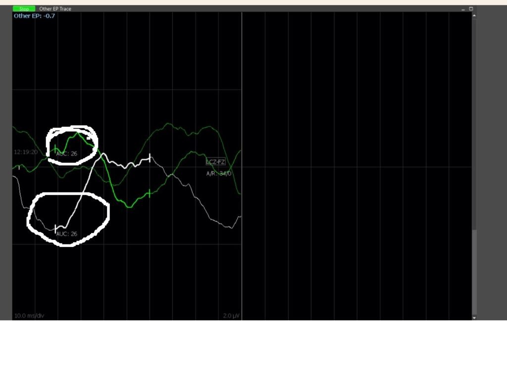
Figure 2 Showing when stimulation applied to the midline in graph top circle is previous findings as a green and bottom circle as a white shows a tiny signal . Its very much important to understand the practical things most of the time we don not get what was written in our text book .
Tumorectomy.
- Based on the information obtained from dorsal column mapping, the surgeon can safely navigate around the sensory pathways to access and remove the intramedullary tumor. By sparing the sensory pathways, the surgeon can minimize the risk of postoperative sensory deficits or neurological complications.
Postoperative Monitoring
- After tumor resection postoperative neurological examination is performed to assess sensory function and ensure that there is no deficits related to the surgery . But in my experience there are mild to moderate sensory deficits because hundred percent preservation of posterior column is difficult still our patient loosing 15-25% sensation here i believe which is acceptable for daily life and further more can be improved .

- Dorsal column mapping is a valuable technique in intramedullary spine tumor surgery for preserving sensory function and minimizing the risk of neurological complications. By accurately mapping out the sensory pathways of the spinal cord, the surgeon can improve the safety and effectiveness of the surgery, leading to better outcomes for the patient.

- What stimulation parameters can be preferred?
- How can be recorded to interpretate the signals?
- Choice of anesthesia drugs ?
- What types of probe can be used?
Related this article.
https://neurointraoperative.com/wp-admin/post.php?post=1794&action=edit
https://www.ncbi.nlm.nih.gov/pmc/articles/PMC10624746
Question.
Which fibers we monitor in mapping?
What can happen if these fiber get injured?
1 thought on “Dorsal column Mapping in intramedullary spine tumor .”