
Reperfusion injury.
Context
Intraoperative neurophysiological monitoring (IONM), particularly TcMEPs, is a key tool during spinal cord surgeries like posterior cervical decompression. It provides real-time feedback about the functional integrity of motor pathways.
In patients with cervical myelopathy, the spinal cord is chronically compressed and ischemic. When decompression is performed, a reperfusion injury may occur, and TcMEPs can show early signs of this.
⚡ TcMEPs: What They Measure
- TcMEPs are recordings of motor pathway responses following electrical stimulation of the motor cortex.
- Reflect descending corticospinal tract function.
- Sensitive to ischemia, injury, or disruption of spinal cord motor pathways.
TcMEP Changes Suggestive of Reperfusion Injury
📉 Sudden Loss or Decrease in Amplitude
- Abrupt drop or loss in TcMEP amplitude immediately after decompression.
- May occur without mechanical cause (no hematoma, no compression on imaging).
🧠 No Change in SSEPs
- Somatosensory evoked potentials (SSEPs) may remain unchanged, supporting a motor-specific spinal cord injury (common in reperfusion injury).
⏱️ Timing
- Changes often occur shortly after decompression when perfusion is restored to chronically ischemic spinal cord tissue.
- There may be a “paradoxical” loss: the patient had stable signals during compression, but signals degrade after decompression.
⚠️ Differential Diagnosis of TcMEP Loss Post-Decompression
| Cause | Features | TcMEPs | SSEPs |
|---|---|---|---|
| Reperfusion injury | After decompression, no mechanical cause | ↓ or lost | Normal |
| Epidural hematoma | May occur later | ↓ or lost | ↓ |
| Vascular compromise (e.g., anterior spinal artery) | May be sudden | ↓ or lost | Normal/↓ |
| Technical issues | Sudden and global changes | ↓ or lost | ↓ or lost |
🧬 Pathophysiology Link
- The ischemic spinal cord is highly vulnerable to free radical and cytokine-induced damage during reperfusion.
- TcMEP loss reflects dysfunction in corticospinal tract neurons, which are particularly susceptible.
🛠️ Clinical Implications
🚨 What to Do Intraoperatively:
- Immediately rule out mechanical causes (check for hematoma, hardware).
- Maintain mean arterial pressure (MAP > 85-90 mmHg) to ensure spinal cord perfusion.
- Administer high-dose steroids (controversial but often considered).
- Neuromonitoring team should document changes, and surgical team may halt or reverse decompression if needed.
🩺 Postoperative:
- MRI to assess for cord edema or infarct.
- Monitor neurologic function closely.
- Initiate rehabilitation early if deficits persist.
📊 Summary Table
| Parameter | Reperfusion Injury |
|---|---|
| Timing | Immediately after decompression |
| TcMEP | Sudden drop or loss |
| SSEP | Usually unchanged |
| Imaging | Cord edema on T2-weighted MRI |
| Management | BP support, steroids, ICU care |
Overview
What is Reperfusion Injury?
Reperfusion injury refers to cellular and tissue damage caused when blood supply returns to the tissue after a period of ischemia (lack of oxygen). The return of blood can cause inflammation and oxidative damage through the production of free radicals.
Pathophysiology in Cervical Myelopathy Surgery
Chronic Compression and Ischemia
- In cervical spondylotic myelopathy, the spinal cord experiences chronic compression, leading to reduced blood flow, metabolic stress, and ischemic changes.
- Over time, the spinal cord adapts to this hypoxic environment.
Decompression and Sudden Reperfusion
- During posterior cervical decompression (e.g., laminectomy, laminoplasty), there’s a sudden restoration of blood flow to previously ischemic regions of the spinal cord.
- This reperfusion can cause:
- Oxidative stress (from reactive oxygen species)
- Inflammation
- Edema
- Disruption of the blood-spinal cord barrier
- Neuronal and glial cell injury
Clinical Presentation
Timing
- Symptoms typically present immediately or within hours after surgery.
Symptoms
- Worsening neurological function after an initially uneventful decompression.
- New or increased motor weakness, sensory deficits, or even paralysis.
- May present with no mechanical cause (e.g., hematoma or hardware compression on imaging).
Diagnosis
Imaging
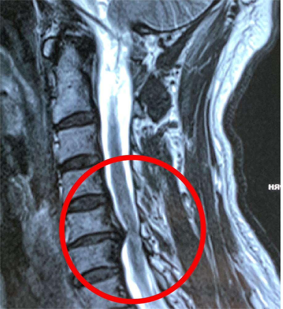
- MRI may show:
- T2 hyperintensity in the spinal cord (suggestive of edema).
- No new compressive lesion.
Rule Out Other Causes
- Rule out surgical complications:
- Epidural hematoma
- Hardware malposition
- Residual compression
Management
Medical Management
- High-dose steroids (e.g., methylprednisolone) may be used to reduce inflammation and edema.
- Close monitoring in an ICU setting.
Supportive Care
- Maintain spinal cord perfusion (adequate blood pressure).
- Physical therapy and rehabilitation if motor function is impaired.
Prevention and Considerations
Preoperative
- Preoperative MRI signal changes (e.g., T2 hyperintensity) can indicate higher risk of reperfusion injury.
Intraoperative
- Gentle decompression techniques.
- Gradual decompression may theoretically reduce risk (though difficult in practice).
Prognosis
- Variable: Some patients may recover completely, while others have permanent neurological deficits.
- Early recognition and treatment are critical to improving outcomes.

https://pmc.ncbi.nlm.nih.gov/articles/PMC11394496
https://neurointraoperative.com/wp-admin/post.php?post=1849&action=edit