
Keywords: Memory Fornix Awake intraoperative mapping White matter stimulation in Third ventricle tumor.
Memory Mapping in Functional Neurosurgery.
Conscious Awake Intraoperative Memory Mapping During Forniceal Stimulation.
The hippocampal complex (HC) is comprised of hippocampus proper the dentate gyrus and the subiculum. The primary efferent fibers from the HC which originate from hippocampus proper and the subiculum, emerge when the HC ends near the splenium of the corpus callosum and become a detached bundle called the crusor ‘leg’ of the fornix. Functionally it has been well established that the HC subserves memory processing (Milner,1970) and lesions to the HC result in deficits of learning and memory (Batchelor, Thompson,& Miller, 2008).
Surgical perspective .
From a neurosurgical perspective manipulation and transection of the fornix is usually avoided. Neurosurgeons have long been taught that disruption of forniceal fibers can result in post operative disability due to declines in memory ,motivation and emotional functioning (Aggletonet et al. 2000, Ehni&Ehni, 1998).
Indication for surgery . for Memory Mapping.
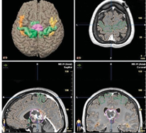

(Scan Image)
White matter tractography image on DTI showing the lesion (magenta) in relation to the left fornix (orange) and the right fornix (yellow).
Memory Mapping .
Awake intraoperative cortical mapping can used direct cortical stimulation while patients perform a variety of cognitive tasks to determine areas of cortex that contribute to language and other cognitive functions so that those areas can be preserved during cortical resection in the neurosurgical treatment of epilepsy or any tumor locating in third ventricle. Subcortical stimulation with bipolar stimulator probe from 6 milliampere and goes up to 12-14 milliamps can utilized to test for the location of the fornices.
The memory testing procedure can be done with visual task like individual line drawn, namable problems to verbally solve (a distractor task).Baseline awake Preoperative memory testing paradigm.
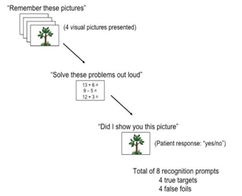
Conscious Awake Intraoperative memory tasks. Each response indicates correct and incorrect .

Conclusion
Awake intraoperative cortical mapping using direct cortical stimulation while patients perform language tasks to assisting with the preservation of cortical areas that subserve language functioning during cortical resection in the neurosurgical treatment of epilepsy or the resection of brain tumors (Haglund et al.,1994; Ojemann et al., 1989).
Awake intraoperative mapping using electrical stimulation to identify white matter areas that are important to cognitive functioning . A paradigm that can be used for awake intraoperative memory testing.
Reference.
Frequently questions can asked .
- Selection of patient for memory mapping. ?
- IONM is useful?
- Purpose of this tool ?
- What types of Anesthesia would preferred?.
- Which part of brain surgery this modality is useful?.
If find this type {Article} useful. Please share and comment down your suggestions.
Related to this article.
https://neurointraoperative.com/wp-admin/post.php?post=1687&action=edit